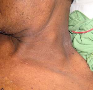
Virchow’s node is a left supraclavicular lymph node. Since there are several nodes in the left supraclavicular region, the enlargement of any one or more of these could be thought of as enlargement of Virchow’s node. The node is also variably termed “Troisier’s node”, “Troisier’s ganglion”, “sentinel node”, and “signal node”. [Free Dictionary, 2013; Casiello, 1958; Scully et al, 2012].
Troisier’s sign is the finding of an enlarged a supraclavicular lymph node caused by an abdominal malignancy.
Historical note: Virchow (pronounced “Firkow” or “Fir-koff”) noted in 1848 that the supraclavicular node became enlarged in gastric cancer. Charles Troisier in 1889 observed that other abdominal cancers too, could enlarge the supraclavicular nodes, hence the eponym “Troisier’s sign”. [Wikipedia]
In the first illustration shown here, one can see a supraclavicular node on the left, and another node higher up in the posterior triangle of the neck. This patient had a colonic cancer. In the illustration below, the patient had a massively enlarged supraclavicular node also from a colonic cancer. Most supraclavicular nodes will be much smaller.
The term “Troisier’s sign” refers specifically to the supraclavicular node that results from an abdominal cancer. This is not to say that the node enlarges only in abdominal cancer. In fact, supraclavicular nodes may be enlarged from any cancer or benign condition that drains into them, including breast cancer [Olivotto et al, 2003] and lung cancer [van Overhagen et al, 2004] and even tuberculosis [Galloy et al, 2005].
Both the right and the left supraclavicular nodes may be involved in disease. Abdominal malignancies metastasize via the thoracic duct, therefore they metastasize exclusively to the left supraclavicular lymph nodes, not the right. Thorax, breast, and head and neck malignancies can go to the neck via the thoracic duct to the left supraclavicular nodes, or via the right lymphatic duct to the right sided nodes. The supraclavicular nodes may also be enlarged in lymphomas and in acute or chronic inflammation, particularly in tuberculosis. However, over 80% of supraclavicular enlargements are malignant.[Cervin et al, 1995]
Pathophysiology
How does the abdominal tumor reach the node?
First, the tumor cells must spread to the lymph in the cisterna chyli, which lies anterior to L1 and L2 vertebrae [Hoit, 2013] The lymph containing tumor cells travels upwards along the thoracic duct, which ends at the venous angle between the left internal jugular vein and the subclavian vein.
Why would the tumor cells move from the duct into the node? There are several hypotheses: [Ludwig, 1991]
- Obstruction causing reflux into the node: if the tumor cells obstruct the duct, the lymph with its constituent cancer cells may reflux into a nearby node that is draining into the duct.
- Reflux during breathing: Reflux may also occur if lymph flows retrogradely due to the negative pressure during respiration.
- Direct flow into nodes: Ludwig [1991] writes that the thoracic duct does not obstruct. He states that supraclavicular node enlargement occurs because, in some patients, the thoracic duct divides into branches that end in lymph nodes. Years earlier Zeidman [1955], too, provided experimental evidence to show that obstruction to the duct was not necessary for chest and neck nodes to receive lymph from the thoracic duct. According to Zeidman the thoracic duct has direct branches to lymph nodes. Later Negus et al [1969] studied supraclavicular nodes by lymphography in volunteers. They noted that lymph flowed into the supraclavicular nodes (numbering 1-24 per person in their study) by reflux, confirming that supraclavicular nodes could receive thoracic duct lymph without obstruction.
Nice descriptions and images are also available in an article by Siosaki and Souza [2014]. and in an article by Loh and Yushak, 2007.
Virchow’s triad, which has nothing to do with Virchow’s node, refers to the three factors that contribute to thrombosis: hypercoagulability, vessel wall defects, and stasis of blood flow.[Scully et al, 2012]
References
- Casiello A. Troisier’s ganglion: pilot sign in the diagnosis of latent stomach cancer. Dia Med. 1958 Jan 6;30(1):10
- Cervin JR, Silverman JF, Loggie BW, Geisinger KR. Virchow’s node revisited. Analysis with clinicopathologic correlation of 152 fine-needle aspiration biopsies of supraclavicular lymph nodes. Arch Pathol Lab Med. 1995;119(8):727-30.
- The Free Dictionary: Troisier, Charles-Emile, accessed 22 Aug 2013
- Galloy AC, Christophe JL, Cambier E, Petein M, Verhelst D. Virchow–Troisier’s node in a haemodialysed patient: it is not always cancer. Nephrol. Dial. Transplant. (2005) 20 (3): 647-649. doi: 10.1093/ndt/gfh543
- Hoit BD. Chylopericardium and cholesterol pericarditis. [UpToDate, http://www.uptodate.com/contents/chylopericardium-and-cholesterol-pericarditis, updated Jul 29, 2013
- Ludwig J. Virchow-Troisier’s lymph node. J Clin Gastroenterol. 1991;13(6):720-1
- Negus D, Edwards JM, Kinmonth JB. Filling of cervical nodes from the thoracic duct and the physiology of Virchow’s node: studies by lymphography. Br J Surg. 1969;56(9):699.
- Olivotto IA, Chua B, Allan SJ, Speers CH, Ragaz J. Long-Term Survival of Patients With Supraclavicular Metastases at Diagnosis of Breast Cancer. J Clin Oncol 2003;21:851-854
- Scully C, Langdon J, Evans J. Marathon of eponyms: 22 Virchow node. Oral Dis 2012;18:107–108
- Loh KY, Yushak AW. Virchow’s Node (Troisier’s Sign). N Engl J Med 2007;357:282, 2007DOI: 10.1056/NEJMicm063871
- Siosaki MD, Souza AT. N Engl J Med 2013; 368:e7February 7, 2013
- Virchow’s node. Wikipedia, accessed 22 Aug 2013
- van Overhagen H, Brakel K, Heijenbrok MW, van Kasteren JHLM, van de Moosdijk CNF, Roldaan AC, van Gils AP, Hansen BE. Metastases in Supraclavicular Lymph Nodes in Lung Cancer: Assessment with Palpation, US, and CT. Radiology 2004; 232:75-80
- Zeidman I. Experimental studies on the spread of cancer in the lymphatic system. III. Tumor emboli in thoracic duct; the pathogenesis of Virchow’s node. Cancer Res. 1955;15(11):719-21


[…] Picture 2. Enlarged lymph node above the left clavicle–Virchow’s node–(the lower of the 2 lumps) in a male with a colonic cancer (Clinipedia) […]
It’s a verry nice notes to go through it…it helped me a lot.
website with tiktok hack
Maulydia had identified Tik Tok Time Hacks back Tik Tok but she however discovered the impression of Tik Tok on someone’s social person when she slogan the crashing on her preteen cousin.